

Read the first chapter
The whole of chapter one, free. About 8 min. Turn the pages with the arrows, your keyboard, or a swipe.
Chapter 1
Why DNA Twists Like a Ladder
The Ladder Hidden in Every Cell
A strand of DNA is only about two nanometres wide, yet it carries the instructions for making a human body, a yeast cell, or an oak tree. Its shape is not a straight line but a twisted ladder, compact enough to fit inside a cell and orderly enough to be copied.
That ladder begins with a simple pairing system. Four chemical bases - adenine, thymine, cytosine, and guanine - join in only two permitted partnerships: adenine with thymine, cytosine with guanine. The pairings hold the two strands together, while their sequence forms a code that cells can read.
The elegance lies in the combination of constraint and variety. The same four letters appear in every human cell, but their order changes from one stretch of DNA to another. A small sequence can help specify a pigment, a protein, or the timing of a cellular event. Across the entire molecule, these sequences become an archive of biological instructions, copied from one generation to the next.
How can four chemical letters carry the astonishing variety of life?
The Twisted Ladder Model
The first useful picture of DNA is not a chain of beads but a ladder turned slowly along its length. The sides are made from alternating sugar and phosphate molecules. The rungs are pairs of bases, each joined to its partner by weak chemical attractions called hydrogen bonds. When the ladder twists, it becomes the double helix.
The pairing rules discovered in the middle of the twentieth century explain why the structure is so well suited to copying. Adenine fits thymine; cytosine fits guanine. Each base on one strand therefore points to the identity of the base on the other. If one strand reads a sequence containing adenine, the matching strand must contain thymine at that position. The molecule carries its own template.
This is the central idea of the Twisted Ladder Model: the sides preserve the molecule’s shape, the rungs store information, and the pairing between rungs allows the information to be reproduced. The model is simple enough to draw on a classroom board, but it describes a mechanism operating inside nearly every living cell.
The two strands also run in opposite directions. This arrangement, known as antiparallel orientation, gives the helix its chemical order and affects how copying enzymes move along it. DNA is not merely twisted for appearance. Its shape is a solution to several practical problems at once: how to store a long message, protect it, read it, and duplicate it.
A single human cell contains roughly two metres of DNA when the molecule is stretched out. It fits inside a nucleus only because it is wrapped around proteins and folded into increasingly compact forms. The result resembles a library whose shelves have been coiled, bundled, and stored in a room too small for the paper to lie flat.
Four Letters, An Endless Archive
The information in DNA does not come from the bases individually. Adenine is not “the eye-colour letter,” and guanine does not carry a fixed meaning by itself. Meaning arises from order.
Cells read DNA in groups of three bases called codons. Each codon corresponds to an amino acid or to a signal that starts or stops the construction of a protein. Because there are four possible bases at each position, a sequence of three positions provides sixty-four possible combinations. Several codons can refer to the same amino acid, a feature that gives the code a degree of built-in flexibility.
Proteins are the working molecules of cells. Some form structures, such as collagen in connective tissue. Others act as enzymes, speeding up chemical reactions, or as signals that help cells communicate. DNA does not build a body directly, like a machine assembled from a single instruction sheet. Instead, it stores sequences that help cells make proteins and regulate when and where those proteins appear.
A gene is best understood as a stretch of DNA involved in producing a functional product, often a protein but sometimes a working RNA molecule. The boundaries and activities of genes are more complicated than the tidy diagrams of early biology suggested. Some DNA helps regulate genes rather than coding for proteins; some regions affect how DNA is folded or read. The ladder is an archive, but it is also a system of switches, bookmarks, and access controls.
This makes heredity less like handing down a finished object and more like passing on a set of construction documents. A child receives DNA from both biological parents, but the instructions are rearranged into a new combination. The child’s cells then read those instructions in the context of development, nutrition, temperature, and countless other conditions.
A surprising fact follows from this: nearly all the cells in a human body contain the same DNA, yet a nerve cell and a liver cell behave very differently. Their difference comes largely from which parts of the shared library they use.
The Discovery of a Shape
The double helix became one of science’s most recognisable images in 1953, when James Watson and Francis Crick published a model of DNA’s structure. Their work depended on evidence gathered by other researchers, including Rosalind Franklin, whose X-ray diffraction images revealed patterns consistent with a helical molecule, and Maurice Wilkins, who also worked with DNA diffraction data.
The history matters because the model was not produced by looking at DNA directly through a microscope. X-ray diffraction scatters X-rays through a crystallised sample, creating a pattern from which molecular structure can be inferred. Franklin’s famous image, often identified as Photo 51, showed a cross-shaped pattern characteristic of a helix. The shape had to be reasoned out from indirect evidence.
Watson and Crick’s model also drew on earlier chemical findings. Erwin Chargaff had observed that the amount of adenine in DNA tended to match the amount of thymine, while guanine matched cytosine. Those relationships made sense once the bases were placed in complementary pairs. The ladder’s rungs were not decorative; they explained the measurements.
The structure solved a biological puzzle that had remained open even after DNA was recognised as hereditary material. A molecule carrying heredity had to do more than exist. It had to preserve information and make copies with enough accuracy for life to continue. Complementary pairing provided a plausible mechanism for both.
Yet copying is not perfectly exact. During DNA replication, cellular machinery checks and repairs many errors, but some changes remain. A base may be substituted, inserted, or deleted. The result is a mutation: a change in the DNA sequence. Some mutations have no noticeable effect, some cause harm, and some alter a trait in ways that may prove useful under particular conditions.
The Surprise in the Copy
The counterintuitive finding is that DNA’s famous stability depends partly on its capacity to change. A molecule that never altered could preserve a lineage, but it could not provide the variation on which evolution acts.
This changes the usual image of genetic accuracy. Copying errors are not simply failures in the system; they are also one source of biological novelty. Their consequences depend on location, context, and environment. A change in a region that does not affect a working instruction may be nearly invisible, while a change in a crucial sequence can alter a protein or disrupt development.
The same principle appears in the spread of antibiotic resistance. In a bacterial population, a mutation may change a cellular target or help a bacterium resist a drug. When the antibiotic removes susceptible competitors, resistant bacteria can become more common. The drug does not deliberately create a useful plan. It changes which existing variations are more likely to persist.
DNA therefore sits between archive and experiment. Its paired bases preserve a message, while occasional copying differences introduce alternatives. Evolution does not write from a blank page. It modifies inherited material, one sequence at a time, under the pressure of changing surroundings.
The Photograph That Made the Helix Visible
At King’s College London in the early 1950s, Rosalind Franklin used X-ray crystallography to study DNA fibres. The work required careful preparation of samples at different humidity levels because DNA changes its shape depending on how much water surrounds it. Her diffraction images recorded patterns too small to see directly but precise enough to reveal the molecule’s geometry.
Photo 51, produced in 1952 with the help of doctoral student Raymond Gosling, showed the distinctive X-shaped diffraction pattern associated with a helix. It was one piece of evidence among several, not a complete picture by itself. The spacing of the pattern helped estimate the distance between repeating units, while other measurements constrained the width and arrangement of the molecule.
The eventual double-helix model connected these observations with Chargaff’s base ratios and the chemical properties of the bases. A structure emerged in which the sugar-phosphate backbones faced outward and the paired bases faced inward. The arrangement explained both the molecule’s strength and its ability to separate into templates during replication.
Franklin died in 1958, four years before Watson, Crick, and Wilkins received the Nobel Prize for work related to the structure of nucleic acids. The episode remains part of the history of DNA not only because of its scientific importance, but because it shows how discoveries are assembled: through instruments, measurements, mathematical interpretation, collaboration, and disputes over recognition.
The image of the helix later escaped the laboratory. It appeared in museums, classrooms, company logos, and public debates about heredity. Its popularity can obscure the physical reality beneath the symbol: DNA is a chemical molecule, continuously copied, folded, repaired, read, and sometimes altered inside living cells.
What the Ladder Leaves Open
The double helix reveals an unusual form of order. Its paired bases make inheritance possible without making life mechanically predictable. A sequence can be copied with remarkable fidelity, yet its effects depend on how cells read it, how other genes interact with it, and what conditions surround the organism.
That tension reaches beyond genetics. Human beings often treat flawlessness as the highest form of design, but living systems persist through a mixture of preservation and variation. The ladder holds its shape by pairing its rungs, while life changes because those rungs are not always reproduced exactly.
A biological instruction is never only a sentence written in four letters; it is a sentence interpreted by a living cell, in a changing world. Somewhere between the stability of the helix and the uncertainty of copying lies the history of every inherited trait - and the question of what the next twist might reveal.
End of chapter one. 7 more chapters in the full book.
Swipe or use the arrows to turn the page


What's inside: 8 chapters
- 1. Why DNA Twists Like a Ladder
- 2. How Cells Copy the Blueprint
- 3. When a Copying Error Helps
- 4. The Traits We Inherit
- 5. Why One Gene Can Change
- 6. Evolution Keeps What Works
- 7. Chance Builds Genetic Diversity
- 8. Imperfection Becomes a Future
About this book
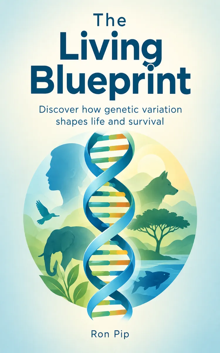
"The Living Blueprint" is a curiosity book by Ron Pip with 8 chapters and approximately 12,347 words. DNA, heredity, mutation, evolution, and the role of variation in survival.
This book was created using Inkfluence AI, an AI-powered book generation platform that helps authors write, design, and publish complete books.
Frequently Asked Questions
What is "The Living Blueprint" about?
DNA, heredity, mutation, evolution, and the role of variation in survival
How many chapters are in "The Living Blueprint"?
The book contains 8 chapters and approximately 12,347 words. Topics covered include Why DNA Twists Like a Ladder, How Cells Copy the Blueprint, When a Copying Error Helps, The Traits We Inherit, and more.
Who wrote "The Living Blueprint"?
This book was written by Ron Pip and created using Inkfluence AI, an AI book generation platform that helps authors write, design, and publish books.
Write your own curiosity book with AI
Describe your idea and Inkfluence writes the whole thing. Free to start.
Start writingCreated with Inkfluence AI


